

Medical treatment for vasospasm consists of “triple-H therapy”: induced hypertension, hemodilution, and hypervolemia which are all aimed at improving cerebral perfusion. Screening of patients at risk for vasospasm is essential as patients cannot be placed prophylactically on treatment for potential vasospasm. High-resistance waveforms will have little to no flow during diastole, whereas low-resistance waveforms will have more abundant forward flow during diastole. The amount of flow during the different portions of the cardiac cycle provides information as to whether the vessel is feeding a high-resistance system such as a muscle bed or extremity or a low-resistance system such as an end organ. Filling in of the spectral window indicates a broader range of flow velocities within the sample volume consistent with turbulent flow seen with stenosis or narrowing (Fig. A narrow spectral line with a clear envelope beneath it indicates fairly uniform (laminar) flow with a narrow range of velocities. The spectral line and the area underneath it (the spectral envelope) can indicate the type of flow present. Again, close attention should be paid to the scale as this can be inverted on the screen. Conventionally, a waveform above the baseline (positive) indicates flow toward the transducer and a waveform below the baseline (negative) indicates flow away. The spectral waveform also indicates directionality of flow. From this waveform, peak systolic velocity (PSV), the velocity of the flowing blood at the peak of systole on the curve, and end-diastolic velocity (EDV), the velocity just before the next systolic upstroke, can be measured. Spectral Doppler allows measurements of flow velocity in a specified area of a vessel (the sample volume) over time and displays these flow velocities as a time/velocity curve throughout the cardiac cycle. In general, the higher the velocity of the blood flow, the greater the degree of stenosis or vasospasm. As elucidated by Spencer and Reid, there is a fairly linear relationship between flow velocity and the degree of stenosis (up to a critical stenosis at which point flow velocities decline due to extreme narrowing of the vessel).

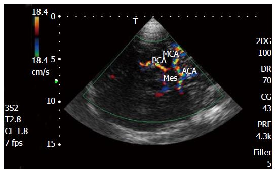
As described above, these flow velocities can be easily calculated by the Doppler equation. The increase in velocity noted in areas of narrowing is a mainstay for diagnosing stenosis or vasospasm in blood vessels. Bernoulli’s principle and the law of conservation of energy also state that the pressure in the region of increased velocity will decrease (Fig. As fluid flows from a conduit (or blood vessel) of greater diameter to one of smaller diameter, the flow velocity must increase to allow the same volume of blood to flow through the narrower area (this is analogous to one putting one’s thumb over the opening of a garden hose as water is flowing out resulting in a high-velocity jet of water being formed). According to the principle, as fluid flows through conduits of differing diameters, the velocity and pressure associated with the fluid will change. At our institution, spectral Doppler waveforms and flow velocities are obtained from the proximal, mid, and distal MCA and single measurements are obtained in the visualized portion of the ACA, PCA, and terminal ICA.īernoulli’s principle, as applied to flowing liquids, can be utilized for the diagnosis of areas of stenosis and vasospasm. With the transtemporal approach, visualization of the MCA, ACA, PCA, and terminal ICA is possible. Flow velocity measurements (both MFV and peak systolic velocity) are obtained. Imaging is performed from the left and right temporal bones with color Doppler flow imaging utilized to visualize the vessels. The transducer is placed on the temporal bone either above the zygomatic arch and anterior to the external auditory canal or slightly more cephalad and posterior over the earlobe. Imaging of the circle of Willis is performed via a transtemporal approach utilizing a 5–1 MHz sector-array transducer with most of the imaging being performed at the lower frequencies (1–2 MHz) to allow penetration of the temporal bone. With advances in ultrasound equipment and the utilization of gray-scale, color Doppler flow, and spectral Doppler imaging, direct visualization of the vessel and more accurate flow velocity measurements are now possible.


 0 kommentar(er)
